ORD Home »
Currents
» New scanning technology could help diagnose Alzheimer's disease using light...
Office of Research & Development |
 |
Office of Research & Development |
|

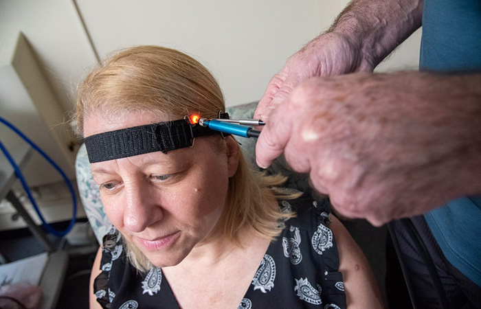
VA researchers at the VA Bedford Healthcare System demonstrate how near-infrared spectroscopy is used to detect brain changes possibly linked to Alzheimer's disease. (Photo by Frank Curran)
July 28, 2021
By Tristan Horrom
VA Research Communications
"This technology is significant because it probes the biochemical and cellular structures of the brain noninvasively with a technique that is inexpensive and could be put into widespread use."
Researchers with the VA Bedford and VA Boston health care systems have developed a non-invasive optical technique to help detect Alzheimer’s disease. The new technique uses spectroscopy—measuring how light is scattered and absorbed when passing through matter—to identify structural changes in the brain.
This scanning method could become a simple, completely non-invasive method of early Alzheimer’s detection, according to the researchers, and also has potential as a way to assess the effectiveness of treatment.
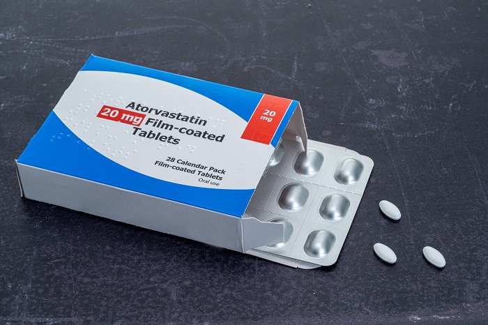
Could cholesterol medicine reduce dementia risk in seniors?

VA study reveals dementia risks unique to people with African ancestry
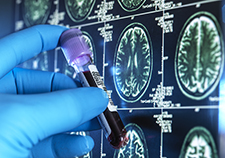
Head trauma, PTSD may increase genetic variant's impact on Alzheimer's risk
“This technology is significant because it probes the biochemical and cellular structures of the brain noninvasively with a technique that is inexpensive and could be put into widespread use, explains Dr. Eugene Hanlon of the VA Bedford Healthcare System, corresponding author on the paper. “Most importantly, it gives useful information about those with mild cognitive impairment.”
The results appear in the June 1, 2021, issue of the Journal of Alzheimer’s Disease.
Alzheimer’s disease is a progressive neurodegenerative disease. It is the leading cause of dementia. Alzheimer’s disease eventually leads to death and no cure currently exists, although medication and supportive treatments can temporarily relieve symptoms.
Alzheimer’s is hard to definitively diagnose at an early stage. Because symptoms are often subtle and gradual at first, the ongoing, irreversible damage is not easy to detect until it is more advanced. Alzheimer’s can definitively be diagnosed only by analyzing brain tissue after death.
While detecting Alzheimer’s disease in a living patient is difficult, imaging technology has made strides toward this goal in recent years. Positron emission tomography (PET) and magnetic resonance imaging (MRI) allow for high resolution images of brain features. These scans can show structural changes related to Alzheimer’s, such as twisted fibers within brain cells and amyloid plaques (misfolded proteins that accumulate between brain cells). They can also detect dynamic features such as how brain cells use glucose.
However, imaging scans such as PET and MRI are expensive, technically demanding, and contribute little to early detection.
VA researchers have developed a new technique that uses light to capture chemical and structural information from brain tissue. The technology works by positioning two fiber-optic probes on the surface of a patient’s temple. One probe delivers near-infrared light non-invasively and harmlessly into the patient’s brain. The other probe collects the light that scatters back.
Near-infrared light—light just at the border of the infrared region of the electromagnetic spectrum—is particularly useful for examining the brain, according to the researchers. It can penetrate deeply into the tissue because the light is only weakly absorbed. This allows for areas of the cerebral to be probed.

Near-infrared light can penetrate deeply into the body’s tissue.
Spectroscopy works by measuring how light moves through and bounces off matter. Different substances block light energy to different degrees, causing the light to be absorbed or scattered. The light is affected at different wavelengths of energy depending on what matter it interacts with. These effects are measured by comparing the light from the source optical fiber with the light collected by the detector fiber. The light detected differs from the initial light because of interactions with the brain tissue.
In collaboration with Boston University’s Alzheimer’s Disease Center, the researchers previously demonstrated the usefulness of this technology using autopsy brain samples from deceased volunteers. Near-infrared spectroscopy was able to distinguish brains confirmed to have Alzheimer’s from those without. By comparing the light refraction from healthy tissue to that of diseased brains, the researchers identified refraction characteristics of tissue affected by Alzheimer’s disease.
In the new study, the researchers applied this technique to three groups of living volunteers: healthy controls, patients with mild cognitive impairment, and late-stage patients who had an Alzheimer’s diagnosis confirmed by autopsy after they died.
They devised a computer algorithm to identify patterns in the spectroscopy data. Through this analysis, the researchers identified two spectral features that signaled the difference between patients with late-stage Alzheimer’s disease from controls with normal brain function. A minor adjustment to those two features allowed the researchers to usefully classify patients with mild cognitive impairment according to degree of impairment. The researchers explain that one spectral feature could be significant in identifying the onset of the disease early on, while the other may be more significant later in the progression of Alzheimer’s. These findings raise the possibility that the method could detect Alzheimer’s disease at an early stage, say the researchers.
This is the first experiment to use such a non-invasive technique to classify a neurodegenerative condition in living patients, according to the researchers.
Beyond helping to identify Alzheimer’s disease, the new technology could also lead to improved treatments, say the researchers. Large clinical trials are still needed to determine whether spectroscopy readings can track disease progression. If they can, explain the researchers, “this approach could become a safe, non-invasive method for assessing response to treatments in real time.”
The new technology could be especially helpful for Veterans. As lead author Dr. Frank Greco explains, “Veterans are more at risk for Alzheimer’s disease than the general population. This technique has the potential to help identify what factors may increase that risk.”
The spectroscopy method has been accepted by the Food and Drug Administration as a protocol for possible clinical use. Before it can be put into practice, clinical trials will need to be conducted. The researchers are working on refining the design of the probe and the specifications of the spectrometer, software, and interpretation of the output toward that end.
VA Research Currents archives || Sign up for VA Research updates