Office of Research & Development |
 |
Talk to the Veterans Crisis Line now
An official website of the United States government


A helicopter gunner watches as the USS Eisenhower passes through the Suez Canal following a deployment in support of Operation Desert Shield in 1990. (DoD photo by Frank Marquart)
July 20, 2017
By Mike Richman
VA Research Communications

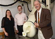
Members of the research team at the Brain Sciences Center at the Minneapolis VA Health Care System, led by Dr. Apostolos Georgopoulos (right). (Photo by April Eilers)
The Brain Sciences Center at the Minneapolis VA Health Care System has evolved into one of the leading facilities in the country for researching Gulf War illness (GWI), a condition that is of major concern to the Veteran community.
Researchers at the center have made what they believe to be key breakthroughs with GWI.
Such is the case with a study, published in June 2017 in Experimental Brain Research, that documents a "systematic and significant" reduction in subcortical brain volumes of about 10 percent in Vets with Gulf War illness, compared with non-affected Veterans. Subcortical is the region of the brain below the cortex, the outer layer of the brain.
Many of the symptoms associated with GWI, especially neurological, cognitive, and mood symptoms, but also fatigue and pain, may be related to the subcortical atrophy observed in the study, according to Dr. Apostolos Georgopoulos, head of the Brain Sciences Center.

This 1995 photo shows Lance Sneath prior to a field worship service he provided for troops during a training exercise with the 2d Infantry Division near the Demilitarized Zone in Korea. (Photo courtesy of L. Sneath)
Former Army chaplain battles brain woes linked to Gulf War illness
In a study that involved 19 Veterans with Gulf War illness, Georgopoulos and his team saw the greatest reductions in the brainstem, cerebellum, and thalamus, and to a lesser extent in the basal ganglia, amygdala, and diencephalon. The highest atrophy was in the brainstem, followed by the left cerebellum and right thalamus, then the right cerebellum and left thalamus.
The brainstem is the part of the base of the brain that is connected to the spinal cord. It controls the flow of messages between the brain and the rest of the body, as well as bodily functions such as breathing, swallowing, heart rate, blood pressure, consciousness, and whether one is awake or sleepy. The cerebellum, which is in the back of the brain, plays a vital role in coordination and balance and affects cognitive functions, such as attention, language, and emotion regulation. The thalamus, which is just above the brain stem, relays motor and sensory signals to the cerebral cortex. It also regulates sleep, alertness, and wakefulness.
Georgopoulos says the impact of the study is "tremendous." The findings "complement and extend" prior research on brain abnormalities associated with GWI and "highlight dramatic and widespread brain effects of Gulf War illness," he says.
"The findings demonstrate that GWI is clearly a brain disorder with impacts on several subcortical structures that are highly interconnected."
"First, the findings demonstrate that GWI is clearly a brain disorder with impacts on several subcortical structures that are highly interconnected," he says. "The specific impacts observed here involved substantial atrophy in several brain areas at a rate that was significantly accelerated compared with healthy controls. Second, by extrapolating the data, we determined that these effects are likely attributable to [exposure to dangerous substances] during the Gulf War period of service."
A 2000 VA-funded report by the U.S. Institute of Medicine (now the National Academy of Medicine) found that Gulf War soldiers were potentially exposed to a wide range of biological and chemical substances. These include sand, smoke from oil-well fires, solvents, insecticides, petroleum fuels, and deadly nerve agents. Many soldiers also consumed pyridostigmine bromide (PB), an anti-nerve agent pill that is alleged to have long-term side effects. (VA has concluded that evidence does not support a link between PB and chronic multi-symptom illnesses in Gulf War Veterans. But VA still presumes that certain medically unexplained illnesses are related to Gulf War service without regard to cause.)
"It is reasonable to suppose that such exposures, alone or in combination, may underlie, in part, the subcortical atrophy observed," Georgopolous says. "We don't know the exact reason and may never know. What we do know is that whatever caused the atrophy has had a lasting impact on the health of these Veterans."
Previous studies have identified brain abnormalities associated with GWI. However, most of that work has focused on specific regions of interest, "potentially obscuring other important effects of Gulf War illness on the brain," Georgopoulos says.
"For instance, abnormalities in the brainstem and basal ganglia have been reported by others," he says. "But aside from our prior brain scans, the cerebellum has received limited attention as it relates to Gulf War illness. Here, we demonstrate that all three areas are substantially affected by Gulf War illness, in addition to several others. Considering brain anatomy, this is not surprising because these regions are highly interconnected."
An estimated 300,000 Veterans have Gulf War illness, which affects various organs, most notably the brain. The condition is a series of medically unexplained chronic symptoms, including fatigue, memory loss, rashes, serious body aches and joint swelling, gastrointestinal problems, depression, anxiety, and headaches.
The study involved 19 Veterans with Gulf War illness (17 men, two women) and with an average age of 48, along with 24 Veterans (23 men, one woman) as controls with an average age of 62. The researchers evaluated brain structure using magnetic resonance imaging (MRI). Previous studies by the Brain Sciences Center on GWI evaluated brain function using magnetoencephalography, a neuroimaging technique for mapping brain activity otherwise known as a MEG scan.
Georgopoulos explains that both methodologies have pointed to brain abnormalities in Gulf War Veterans, with the MEG scan identifying cortical anomalies in neural communication, particularly involving the cerebellum. Neural communication is the signaling between neurons in the nervous system. The MRIs showed atrophy in the cerebellum and other closely linked areas, such as the brainstem and thalamus, he says.
The MRI testing found "highly significant reductions" in subcortical brain volume in the Gulf War illness participants, compared with the controls. Those with GWI showed reductions of 12.3 percent in the brainstem, 10.4 percent in the cerebellum, and 9.7 percent in the thalamus, relative to the controls.
To determine the point when the brain atrophy may have started, the researchers extrapolated data to learn when the lines from the GWI participants and controls intersected. They compared that information to the age of the participants on Jan. 1, 1991, the same month U.S. forces entered the war.
For the cerebellum, the lines intersected at age 28, which was well within the range of the participants' ages when they were activated for the Gulf War. This meant those soldiers began losing some of the cerebellum at a rate much faster than the controls. In comparison, the lines intersected at age 36 for the brainstem, which was out of the range of war-time activation. The researchers thus concluded that the brainstem atrophy was likely secondary to the cerebellar atrophy.
However, Georgopoulos says the deficit in the cerebellum impacted the brainstem, noting that a large part of brainstem volume is related to the cerebellum. Major systems of the cerebellum are located in or pass through the brainstem. In contrast, a relatively small fraction of the thalamus is related to the cerebellum, he notes.
Studies have linked the deterioration of nerve cells in the cerebellum to autoimmune disorders such as multiple sclerosis and Hashimoto's thyroiditis. "It seems that the cerebellum is a frequent target organ of such disorders, as well as of dangerous toxins, the researchers write.
They say the research dovetails with a prior study at the Brain Sciences Center that showed anomalies with neural communication, mainly in the cerebellum. In that study, the scientists documented sharp differences in brain function between healthy and ill Gulf War Veterans in the cerebellum and frontal cortex. The researchers found distinctions between the two groups in synchronous neural interactions, also known as synchrony. Such differences are excellent biomarkers of GWI, say the researchers.
Furthermore, the atrophy in the brainstem, cerebellum, and thalamus, together with the reductions in size in the basal ganglia, amygdala, and diencephalon, resemble the distribution of brain atrophy that's been linked to toxic encephalopathy, the researchers say. Toxic encephalopathy refers mostly to abnormalities in the water, air, and other organic solvents that affect brain function.
Many diseases that cause brain atrophy have been associated with dementia and other degenerative brain diseases. But Georgopoulos stops short of saying the subcortical brain atrophy identified in his latest study will lead to one of those conditions.
"Given the myriad interconnections in the brain, it is safe to say that accelerated atrophy will ultimately impact functioning," he says. "However, the specific effects are determined by a number of factors, including the location of atrophy and the rate of atrophy."
Georgopoulos notes that the Brain Sciences Center is planning future studies that may provide insight into the exact cause of the subcortical brain atrophy observed in its most recent research. Those studies may also offer insight into potential treatment options that could help limit brain atrophy in Vets with Gulf War illness, he says.
This 1995 photo shows Lance Sneath prior to a field worship service he provided for troops during a training exercise with the 2d Infantry Division near the Demilitarized Zone in Korea. (Photo courtesy of L. Sneath)
Lance Sneath served as an Army chaplain for more than two decades. He counseled soldiers while on deployment during the Gulf War and supported service members and families in the pre- and post-9/11 era. He comforted those who grieved for troops killed in Iraq and Afghanistan and helped others whose loved ones were prisoners of war.
But Sneath can no longer offer that emotional support. He's battling a myriad of conditions that are hallmarks of Gulf War illness and have derailed his personal and professional life.
His brain has deteriorated to the point where he has seizures and cognitive problems such as short-term memory loss and concentration lapses.
He has three autoimmune disorders that are believed to cause brain abnormalities: multiple sclerosis, a nervous system disease that disrupts the flow of information within the brain, and between the brain and body; Hashimoto's thyroiditis, in which antibodies attack the thyroid, leading to a decline in that gland's ability to function; and celiac disease, in which the ingestion of gluten can lead to intestinal damage.
Plus, he has fibromyalgia, a disorder characterized by widespread musculoskeletal pain accompanied by fatigue, sleep, memory, and mood issues.
Sneath, 55, says his doctor told him when he was 50 that his brain had atrophied, or declined in size due to a loss of cells, to where he was functioning with a 75-year-old brain. Brain atrophy is a key sign of Alzheimer's disease, a progressive brain disorder that destroys memory, thinking skills, and the ability to carry out simple tasks.
"I had a great mind that one day I lost," says Sneath, who has two master's degrees, one in divinity and another in counseling psychology, and has published articles on counseling military families.
"I drove to my family life training center one morning, but things were very different. It was like a fog came into my mind. I knew basically who I was, but I didn't know what I was doing at the center. I walked into the center, and some of these people who I'd known for the last 10 or 15 years by their first names, I couldn't remember any of them. It was like I'd lost my mind on the way to work. To be at the top of your professional game, so to speak, and suddenly just have everything taken away..."
Like many other Gulf War Vets, Sneath believes he was exposed to deadly chemical agents and other toxins during the war. In January 1991, he was based in Dhahran, Saudi Arabia, as part of the Army XVIII Airborne Corps when a U.S. missile intercepted and destroyed an Iraqi Scud missile that was targeted at the Saudis. His superiors said the Scud contained no deadly toxins, but he has since read that traces of sarin nerve gas may have descended on his camp in a light mist after the Scud exploded.
Sneath thinks he had a similar exposure when a Scud exploded in the skies near the Saudi city of Hafar Al-Batin, another base of his during the war. He also recalls being in the vicinity of burning oil fields with huge plumes of smoke and "horrific" sandstorms in Iraq, noting that the sand particles could have been carrying toxins.
These days, Sneath takes 23 daily medications and sleeps an average of 18 hours per day. "If I get a good six hours out of bed during the day, I count it a real victory," he says. Having served four years in the Air Force in the 1980s, he often sees physicians at Sheppard Air Force Base in Texas.
Recently, he visited the Brain Sciences Center at the Minneapolis VA and underwent two brain imaging procedures: magnetic resonance imaging and a MEG scan. He also had cognitive testing that confirmed his short-term memory is "pretty bad," he says.
Dr. Brian Engdahl, a psychologist at the Brain Sciences Center, says Sneath's exposures to deadly toxins may have contributed to his autoimmune disorders and his body's failure to clear itself of pathogens and poisons.
"The autoimmune responses are attacking the brain and impairing the ability of many Gulf War Veterans like Lance to think and concentrate," Engdahl says. "Many of these Veterans are experiencing memory and mood problems."
In learning about the research at the Brain Sciences Center, Sneath feels there's hope for a treatment with the potential to improve the brain-related functions that he and many other Gulf War Vets have lost. He's particularly interested in the center's research that identified brain mechanisms and other areas involved in Gulf War illness (GWI). The research found that certain forms, or alleles, of HLA (human leukocyte antigen) genes offer protection from GWI. HLA genes play key roles in immune system functioning, but a lack of the alleles has made Veterans vulnerable to developing GWI symptoms.
"My worst fear is being forgotten and [left] on my own to live with this nightmare," Sneath says. "Going to the Brain Sciences Center reassured me and my wife that we not alone in this battle!"
VA Research Currents archives || Sign up for VA Research updates