Office of Research & Development |
 |
Talk to the Veterans Crisis Line now
An official website of the United States government

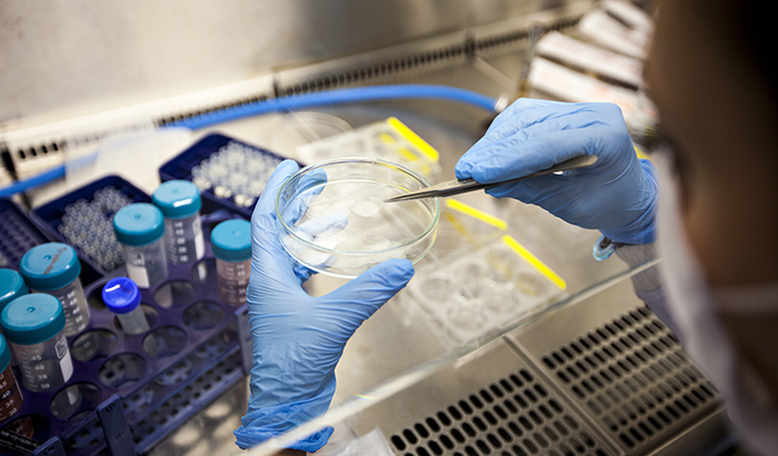
Scientists make brain organoids by culturing human stem cells. They use them to study a range of brain diseases. (Photo: ©iStock/gevende)
April 23, 2020
By Mike Richman
VA Research Communications
"When we're trying to understand human brain diseases, obviously there's not as much we can do with the human brain itself."
Traumatic brain injury, Parkinson’s disease, schizophrenia, stroke—there’s no shortage of research on diagnosing and treating these and many other brain conditions.
But one approach you may not have heard of yet involves human brain organoids. The experimental technology promises to transform how brain diseases are understood and treated.
Also known as cerebral organoids, these miniature structures are simplified versions of areas of the human brain. Scientists make them by culturing human stem cells in a 3D rotational bioreactor and use them as models to better understand brain diseases. The models are in the earliest stages of development.
Brain organoids are central to the research of Dr. H. Isaac Chen, a neurosurgeon at the Corporal Michael J. Crescenz Veterans Affairs Medical Center in Philadelphia. He led a review study on the topic that appeared in the journal Developmental Dynamics in January 2019.

Blood pressure drug could prevent posttraumatic headaches

Five ways the Million Veteran Program is transforming our understanding of Veteran health

Traumatic brain injury carries risk for cardiovascular disease in post-9/11 Veterans
Head trauma, PTSD may increase genetic variant's impact on Alzheimer's risk
“Brain organoids have inspired such intense excitement over the past five years because they are the only [lab-based] platform that recreates the 3D architecture of the human brain and [repeats] the process of human neurodevelopment,” Chen and his colleagues write. Neurodevelopment refers to the formation of the brain.
The researchers add: “Brain organoids are human systems that better match the structural background and genetic characteristics of the human brain than animal models. They are also much more accessible and available in greater quantities than human brain tissue. Lastly, organoids are 3d structures that [repeat] many aspects of the [real-life] environment of brain tissue. Such features are missing in two-dimensional [one cell thick] cultures.”
In the human brain, ventricles are fluid-filled structures in the center of the brain. They are surrounded by stem cells that generate neurons. “This arrangement results in layers of neurons on top of the stem cells, which are themselves on top of the ventricle,” says Chen, who is also an assistant professor at the University of Pennsylvania. “This same structure is seen in brain organoids.”
Currently, Chen explains, scientists are creating organoids mostly to replicate specific areas of the brain, such as the cerebral cortex, the hippocampus, and the pituitary gland. The most common area being studied is the cerebral cortex, a large part of the brain that plays a key role in consciousness. Many of the motor or cognitive functions that are lost from traumatic brain injury, for example, are related to the cerebral cortex in “some way, shape, or form,” Chen says, adding that a stroke can also impact the cerebral cortex.
Researchers are also using organoids to recreate areas of the brain that produce dopamine, such as the midbrain, for the purposes of studying Parkinson’s disease, a neurological disorder that affects movement. Research has shown that dopamine, which plays a central role in the “reward system” of the brain, is present in parts of the midbrain. A deficiency of dopamine means movements may become delayed and uncoordinated.
“When we’re trying to understand human brain diseases, obviously there’s not as much we can do with the human brain itself,” Chen says. “We have to rely on models. Commonly, animal models are used. But there are major differences between the human brain and that of a rat or pig or monkey. So we often end up with results that may not be directly generalizable to the human condition.
“Especially in cases of the central nervous system, the brain, traumatic brain injury, or stroke, many therapies have worked in animals,” he adds. “But when efforts are made to translate them to humans, they don’t work. One thought is that this is due to the differences between brain cellular or molecular structure between a rat versus a human. So if you have something that’s a little closer to the human brain, such as a brain organoid, then you might be able to get some insight into a disease that actually has much more relevance to human conditions.”
In the review, Chen and his colleagues focused on three diseases—the Zika virus, autism, and Glioblastoma multiforme–to consider how organoids could contribute to disease modeling, personalized medicine, and the testing of therapeutics.
The Zika virus is a mosquito-borne virus that can increase the risk of neurological problems, particularly an abnormally small head in babies born to infected mothers. Autism is a group of developmental disorders that can involve language and social impairments and repetitive behaviors. Glioblastoma multiforme, a common form of a brain tumor in adults, is one of the hardest cancers to treat, with a median life expectancy of only 15 months.
Brain organoids, according to the researchers, helped show that the Zika virus led to a loss of cells that produce neurons, resulting in a smaller brain.
On autism spectrum disorder (ASD), “It stands to reason that human models are most suitable for studying ASD because of the involvement of the cerebral cortex, which is more highly developed in humans than other species, in ASD pathology,” the researchers write. “Moreover, ASD pathology is likely related to abnormalities in how different parts of the brain are connected to each other. Thus, ASD models ideally should have networks of neurons that appropriately simulate brain circuitry. Brain organoids fulfill these criteria.”
Regarding glioblastoma multiforme, the research team explained that organoid technology can be used to study brain tumors in two ways.
“First, the same techniques that give rise to brain organoids can be applied to brain tumor tissue taken from patients, resulting in `glioma organoids,’” Chen says. “These organoids can be used to study the biology of brain organoids in the laboratory in three dimensions without needing an animal host. Glioma organoids or brain tumor cells can also be fused to normal organoids to study how brain tumor cells invade the brain, one of the hardest problems to address with brain tumors.”
Chen and his team also discussed improvements needed in brain organoids to enhance their role in research, as well as their potential role in repairing brain circuits after injury. Scientists need to make a lot of high-quality organoids, improve them so they model the brain as closely as possible, and better understand the cellular pathways in a disease, Chen says.
“The problem is not needing enough organoids to model an area of the brain like the cerebral cortex,” he says. “It’s that you need multiple organoid samples to conduct studies and achieve enough statistical power to see real differences between the groups.”
Additionally, Chen and his team acknowledge that brain organoids invite “potential ethical quandaries.” There are theoretical concerns, Chen says, that organoids could develop brain functions, such as the sensation of pain or awareness of the environment, while they are being cultured in a lab. “If these features arise, then organoids may need additional protections, such as those given to animals that are involved in research,” he says. “For example, if an organoid could feel pain, measures would be needed to minimize the pain that it experiences. However, this possibility is highly unlikely given the current small size of brain organoids and their limited neural activity.”
As models evolve to resemble the human brain more closely, concerns will also exist about what it means for human organoids to be transplanted into the brain of an animal.
“Would it be possible for the rat to become smarter?” Chen asks. “There have been decades of discussion on potential outcomes of putting human neurons in animal brains, and brain organoid transplantation falls within this discussion. But local transplantation of brain organoids is unlikely to improve an animal’s brain function also because of their small size and limited neural activity. Scientists would need to repair brain function first before being able to enhance an animal’s brain. Nonetheless, careful behavioral testing of animals is needed as these studies progress, and animal care with regard to pain and discomfort needs to adhere to currently in-place guidelines for animal research.”
Chen believes brain organoids are 5 to 10 years away from being tested in human clinical trials that focus on brain repair. Animal studies on this are in the early stages.
“Much needs to be done before brain organoids become a part of standard medical practice,” he says. “A lot of this has to do with improving the structure and composition of brain organoids. The neural activity of brain organoids is also just beginning to be understood. These issues will require quite a bit of further study before organoids can routinely be used for modeling brain diseases and disorders and as tissues for brain repair.”
VA Research Currents archives || Sign up for VA Research updates